|
Semi-automatic segmentation of 3D ultrasound images of the
prostate |
|
|
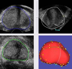
This project aims at facilitating the segmentation of the prostate in
ultrasound 3D images using shape priors. An average prostate coming from a statistical atlas is positioned
in the ultrasound volume and deformed to fit to user-defined points of the
prostate contours. Generally, thirty to forty points are sufficient to
guarantee a precise result. Where 20 to 30 minutes were necessary in a
completely manual segmentation, a similarly good segmentation can be obtained
in five minutes with this method. |
|
|
Reference: K. Dowlut,
M. Baumann, S. Martin, J. Troccaz - Semi-automatic
segmentation of prostate ultrasound images using shape priors - Poster
presented to: Engineering and Urology Society 24th Annual
Meeting, Chicago, 25 Avril 2009 – on-line
proceedings freely available |
|
|
Collaborations: Clinical
partners and KOELIS company. |
|
|
Status: FINISHED This project was developed by Kevin Dowlut during a training session of the « Magistère informatique” de l’UJF (Grenoble University). The developed tools have
been integrated in the Urostation platform of
KOELIS. |
|
|
Contact : jocelyne.troccaz@imag.fr |
|