|
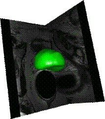
Segmentation using a statistical atlas (application to prostate MRI) |
|
|
The
objective is to use a priori information embedded in a model for automatic
segmentation of 3D volumes (e.g. MRI data). Based on existing images segmented
by an expert operator, an atlas is built after elastic registration of both
the exams and their associated segmentations. The atlas includes the average
image, the contours “projected” to this average image and a statistical model
of the prostate surface. Segmenting a new patient requires the registration
of the exam to the atlas. After registration contours and statistical data
are used for segmentation of the new exam. Though the approach is based on a
hybrid registration combining feature-based and image-based frameworks. |
|
|
References: S. Martin, V. Daanen, J. Troccaz. Automated Segmentation of the Prostate in 3D MR Images Using a Probabilistic Atlas and a Spatially Constrained Deformable Model, Medical Physics, 2010, 37(4) :1579-1590 (pdf ) |
|
|
Collaborations : CHU de
Grenoble (Dr Philippe Fourneret – Radiotherapy department) |
|
|
The
developed segmentation tools have been integrated to the Urostation
from KOELIS. |
|