|
AI-based Automatic segmentation of 3D ultrasound images of
the prostate |
|
|
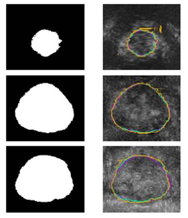
The objective is to produce automatically contours of the prostate
from 3D ultrasound images. The 3D volume is represented
as a stack of 2D slices. The approach is based on a combination of convolutional
networks: 3 networks specialize in the segmentation of 2D slices in a given
direction (axial, sagittal, frontal) while the 4th
network develops an ensemble learning strategy; for each voxel of the volume,
it determines the optimal weighted combination of the three segmentations by
the direction-specific networks. The rationale is that some regions of the
organ are more visible on a specific viewing direction (for instance the apex
of the prostate is more visible on sagittal slices). The method has been trained and evaluated on a clinical database from the
Grenoble University Hospital (in agreement with French regulation on clinical
data). |
|
|
Reference: Clément Beitone and
Jocelyne Troccaz. Multi-expert fusion: An ensemble learning framework to
segment 3D TRUS prostate images. Medical Physics, 2022:49(8):5138-48. https://hal.archives-ouvertes.fr/hal-03654488 . |
|
|
Funding: Region “Rhône-Alpes
Auvergne” project PRONAVIA, PSPC DIANA
from BPI France and MIAI AI Institute |
|
|
Collaborations: Clinical partners
and KOELIS company. |
|
|
Status: IN
PROGRESS Work with Clément BEITONE, TIMC |
|
|
Contact : jocelyne.troccaz@univ-grenoble-alpes.fr and clement.beitone@univ-grenoble-alpes.fr |
|