|
Automatic segmentation of brachytherapy seeds in 3D
ultrasound images |
|
|
In order to allow
dynamic and adaptive, robotic or manual brachytherapy, it is essential to be
able to robustly detect the real position of the deposited
grains. In addition to the intrinsic difficulties linked to segmentation in
ultrasound imaging, there are the specificities linked to the grains
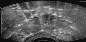
(smallness of the grains and very many artefacts they produce - see opposite
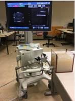
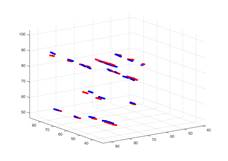
above). We have developed an approach using a prior detection of needles via a Bayesian classification method to infer a search area. The grains are detected thanks to the registration of a geometric model of the grain train with the image segmented by Bayesian classification in the search area. Then for each grain, the position is then refined by iconic registration of a grain appearance model in the images with the sub-volume concerned. This approach has been evaluated on phantoms (see opposite, bottom) and on patient data. |
|
|
References : Hatem Younes, Jocelyne Troccaz, and Sandrine Voros. Machine learning and registration for automatic
seed localization in 3D US images for prostate brachytherapy. Medical Physics, 48(3):1144-1156, January 2021. https://hal.archives-ouvertes.fr/hal-03023560 . Younes, H., Voros, S., & Troccaz, J. (2018,
April). Automatic needle localization in 3D ultrasound images for
brachytherapy. In 2018 IEEE 15th International Symposium on Biomedical
Imaging (ISBI 2018) (pp. 1203-1207). IEEE. https://hal.archives-ouvertes.fr/hal-01800108/document PhD
thesis of Hatem Younes, 2020, in French (https://hal-emse.ccsd.cnrs.fr/INPG/tel-03009135v1
) |
|
|
Funding: ANR FOCUS project (coordinated by LATIM, Brest,
France) |
|
|
Collaborations : Partners
of the FOCUS project (LATIM lab, Brest University Hospital, Grenoble
University Hospital, Koelis company) |
|
|
Status : FINISHED |
|
|
Contact : jocelyne.troccaz@univ-grenoble-alpes.fr
|
|