|
Biomechanical modelling of the prostate |
|
|
|
Despite the important number of algorithms proposed
for interactively modelling deformable objects such as soft human tissues, very
few methods have attempted to simulate complex anatomical configurations
taking care of both of interactivity and realism in terms of predictions of
tissue motions and deformations. For several years, we have worked on the
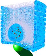
development and evaluation/validation of biomechanical prostate models
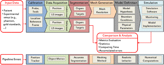
clinically usable. We have recently proposed a full pipeline for the study
and evaluation of image-based biomechanical models. It has been successfully
applied to modelling the prostate deformations in the context of ultrasound
guided interventions (biopsy for instance). |
|
Reference: Sarrazin, J. (2016). Modélisation
biomécanique pour l'imagerie de la prostate. Thèse de Doctorat, université de
Grenoble Alpes. (pdf) |
|
Status: ACTIVE PhD theses of M.Marchal in 2006 and J.Sarrazin in 2016. Funding through the ANR Prosbot and FUI MIRAS projects. Collaboration with Koelis. |
|
Contact :
Emmanuel.Promayon@imag.fr or jocelyne.troccaz@imag.fr |