|
MRI/histology registration. Application to prostate
cancer |
|
|
Histology is the gold standard for cancer diagnosis, grading and
localization. We have developed the ProPath system
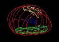
enabling 3D reconstruction of a model from histologic
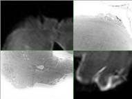
slices and registration to MRI data. The clinical application is prostate
cancer. Reconstruction is based on the Stanford protocol. Registration is a
3D/3D elastic surface registration. One objective is a better understanding
of MRI visualisation of cancers. A longer term objective is to build a
statistical map of prostate cancer occurrence in the different anatomical
zones of the prostate in order to assist biopsy planning. |
|
|
Reference: Bart S., Mozer P., Hemar P., Lenaour G., Comperat E., Renard-Penna R., Chartier-Kastler
E., Troccaz J. MRI-histology registration in
prostate cancer. In Proceedings of Surgetica’2005, Merloz P.,
Troccaz J. (Eds), Sauramps
Medical, 2005, pp361-367 (PDF) |
|
|
Collaborations: La Pitié Salpétrière Hospital, Paris (Dr
Bart, Dr Mozer, Dr Chartier-Kastler – urology dept –anatomopathology dept – imaging dept) |
|
|
Status: FINISHED Experimental work done on specimen organs. This
project was funded by PHRC national 2003 « Prostate-Echo » |
|
|
Contact: jocelyne.troccaz@imag.fr |
|