MRI /US data fusion for prostate brachytherapy

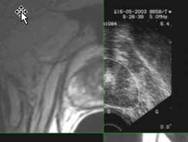
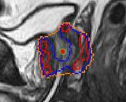
This project aimed at improving echographic data interpretation during prostate brachytherapy. Pre-operative MRI data are automatically registered to intra-operative trans-rectal ultrasound images using a 3D/3D elastic surface matching. For each echographic image, the corresponding MRI data are computed and a composite image is produced. The user can interact with the image. This facilitates US image segmentation (especially close to the prostate extremities) from which dosimetric planning is performed. We compared the dosimetry performed on the original US images to the one that would be obtained on MRI-enhanced US images. It demonstrated a systematic under-estimate of the prostatic volume and over-estimate of the delivered dose when MRI is not used. This study involved 28 patients. A new version of the approach has been developed based on the use of MR contours for US automatic segmentation.

image-composite

close-up-sag2

References :

Martin S., Baumann M., Daanen V., Troccaz J. MR prior based automatic segmentation of the prostate in TRUS images for MR/TRUS data fusion, IEEE International Symposium on Biomedical Imaging, ISBI’2010, Rotterdam, 14-17 Avril 2010, pp640-643 (pdf

)

V. Daanen, J. Gastaldo, J.Y. Giraud, P. Fourneret, J.L. Descotes, M. Bolla, D. Collomb, J. Troccaz. MRI/TRUS data fusion for brachytherapy. The International Journal of Medical Robotics and Computer-Assisted Surgery, Vol2, No.3, pp256-261, September 2006

Collaborations: Grenoble University Hospital (Pr Descotes – Urology department; Dr Fourneret, Pr Bolla and Mr Giraud, Radiotherapy dept) – KOELIS company

Status: FINISHED

Application validated on patients (cf. Supra) and new approach validated on patient data. MRI/US registration has been integrated in the Urostation of KOELIS.

Contact: jocelyne.troccaz@imag.fr or vincent.daanen@imag.fr