|
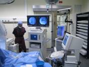
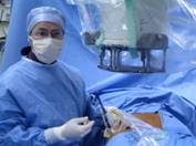
Fluoronavigation of US-guided renal punctures |
|
|
Traditionally,
renal punctures are performed using either X-ray images which clearly show
the renal cavities whilst being invasive or ultrasound images which are not invasive
but have a limited readability. To avoid a complex registration approach for
combining both modalities we have participated to the development and
evaluation of an alternative approach based on fluoronavigation.
This approach couples X-ray and ultrasound data in a very simple way using a
localizer. After having recorded calibrated X-ray images, the puncture
trajectory materialized by a mechanical guide attached to the Though
the operator can benefit from the advantages of both modalities when
performing the puncture. |
|
|
Reference: P. Mozer, P. Conort, A. Leroy, M.
Baumann, Y. Payan, J. Troccaz,
E. Chartier-Kastler, F. Richard. Aid to percutaneous renal access by virtual projection of the
ultrasound puncture tract onto fluoroscopic images. J Endourol. 2007
May;21(5):460-465 |
|
|
Collaborations : Koelis
company – |
|
|
Status : FINISHED EC marked system distributed by the KOELIS company |
|