|
3D reconstruction of biopsy series |
|
|
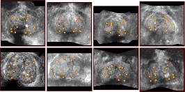
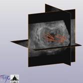
Traditionally prostate biopsy series (for instance 12 samples taken
out of the gland as regularly as possible) are executed under endorectal 2D ultrasound control. The real position of
the samples is never exactly known. Based on 3D ultrasound elastic
registration tools, we developed a method for accurate localization of the
biopsies in a single referential. Such a representation enables precise
localization of tumoral regions of the gland if any
detected by anatomo-pathologists and targeted
treatments. A clinical study involving more than 50 patients has demonstrated
quantitatively the systematic lack of samples for some regions of the
prostate and a learning curve due to the 3D representation of the biopsy
sessions to the clinician. This system also enables mapping several biopsy
series of a same patient performed at different times. |
|
|
Reference : P.Mozer. « Urologie et GMCAO » (in
french), Ph.D thesis, UJF, Juin 2007 (pdf can be downloaded from the HAL database) P. Mozer, M. Baumann, G. Chevreau, A. Moreau-Gaudry, S. Bart, R. Renard-Penna, E. Comperat, P. Conort, M.-O. Bitker, E. Chartier-Kastler, F. Richard, J. Troccaz. Mapping of Transrectal Ultrasound Prostate Biopsies: Quality Control and Learning Curve Assessment by Image Processing, Journal of Ultrasound in Medicine, 2009, 28(4):455-460 460 (pdf |
|
|
Collaborations : Urology department of La Pitié Salpétrière
Hospital (Dr Mozer) – société KOELIS |
|
|
More
information: Best thesis award 2008
SFGBM/IEEE EMBS (section France) |
|
|
Contact : jocelyne.troccaz@imag.fr or pierre.mozer@psl.aphp.fr |
|