|
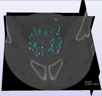
Seed detection in CT scans for prostate
brachytherapy |
|
|
Prostate brachytherapy generally involves a
post-implantation CT acquisition (one month later) for a dose estimation
relying on real position of implants which may
differ from planned ones. On-the-shelf software generally assumes that the
seeds are aligned with the implantation direction. Moreover, cluster of seeds
may lead to false positive or negative detections. We have developed a new
method enabling the localization of seeds (5 parameters since seeds are
cylinders) and the ability to handle seed clusters. The method is automatic,
reliable and accurate as demonstrated on data from phantoms and 16 patients. In order to evaluate the approach on a large scale,
a new version of the software for clinical users, coupled with dose
computation, has been developed. |
|
|
References : N’Guyen H-G, Fouard C, Troccaz J. Segmentation,
separation and pose estimation of prostate brachytherapy seeds in CT images,
IEEE Transactions on Biomedical Engineering, on line first 6 March 2015 (pdf) N’Guyen G, Fouard C, Meneu
F, Giraud JY, Troccaz J. Automatic 3D seed location and orientation detection
in CT images for prostate brachytherapy. Proceedings of IEEE ISBI’2014,
pp1320-1323, Beijing, 28 April-2 May 2014 (pdf) |
|
|
Collaborations: radiation oncology department (JY Giraud, C Verri)
and LPSC lab for Dorgipro project. |
|
|
Funding: INSERM Dorgipro project and « coup de pouce »
ECCAMI. |
|
|
Status :
ACTIVE Postdoc of Huu-Giao N’Guyen
in collaboration with P.Chevalier and F.Meneu (master student medical physics). Clinical
application transfer development by Jean-Loup Haberbusch
(ECCAMI). Master of Kumar Kumar Tamizhanban, radiation
therapy resident, CHU Grenoble. |
|
|
Contact :
jocelyne.troccaz@univ-grenoble-alpes.fr
|
|